Novel chemical-physical autopsy investigation in sudden infant death and sudden intrauterine unexplained death syndromes
Antonietta M Gatti, Marko Ristic, Stefano Stanzani & Anna M Lavezzi
Published Online:8 Feb 2022https://doi.org/10.2217/nnm-2021-0203
Abstract
Aim: Verify the presence of inorganic nanoparticle entities in brain tissue samples from sudden infant death syndrome (SIDS)/sudden intrauterine unexplained death syndrome (SIUDS) cases. The presence of inorganic debris could be a cofactor that compromises proper brain tissue functionality. Materials & methods: A novel autopsy approach that consists of neuropathological analysis procedures combined with energy dispersive spectroscopy/field emission gun environmental scanning electron microscopy investigations was implemented on 10 SIDS/SIUDS cases, whereas control samples were obtained from 10 cases of fetal/infant death from known cause. Results: Developmental abnormalities of the brain were associated with the presence of foreign bodies. Although nanoparticles were present as well in control samples, they were not associated with histological brain anomalies, as was the case in SIDS/SIUDS. Conclusion: Inorganic particles present in brain tissues demonstrate their ability to cross the hemato–encephalic barrier and to interact with tissues and cells in an unknown yet pathological fashion. This gives a rationale to consider them as cofactors of lethality.
Sudden intrauterine unexplained death syndrome (SIUDS) and sudden infant death syndrome (SIDS) are unresolved social and health problems even today [1–3]. There are no specific pathognomonic or diagnostic findings identified so far at the routine autopsy of SIUDS and SIDS victims. Cause of death remains largely unexplained; however, in-depth brain examination has increasingly disclosed a variety of developmental abnormalities of brain centers that are crucial for the control of autonomic and cardiorespiratory functions. Such structures include the Kölliker-Fuse nucleus, the facial/parafacial complex in the pons and the pre-Bötzinger nucleus in the medulla oblongata [4–6]. Defective synthesis and deregulated expression of various neurochemicals (catecholamines, serotonin, somatostatin, orexin, nicotinic acetylcholine receptors, growth factors etc.) are frequently found in both SIUDS and SIDS [7–11]. However, morphological and functional alterations highlighted in SIUDS and SIDS are frequently not sufficient to explain their etiopathogenesis. It is increasingly clear that sudden fetus or infant deaths are due to multiple factors. Before the modern age of metabolic diseases, death was caused mostly by environmental events, infection, pollution or disasters. Today, in the Pandora's box of environmental pollutants influencing human well-being, one would need to add nano-sized environmental pollutants (NEPs), whose abundance and significance increased dramatically in the past few years.
‼️‼️‼️‼️‼️‼️‼️‼️‼️‼️
NEPs derived from nanomaterials used in biomedicine, biotechnology and environmental industry [12,13] are known to cross the blood–brain barrier [14]. As metal nanoparticles have already been found in fetal kidney and liver tissues [15], it is easy to suspect that if taken up by a pregnant woman ‼️(through inhalation, ingestion, injection, skin contact etc.)‼️, NEPs can enter the mother's blood, easily cross the placental barrier and finally enter the fetal bloodstream [16]. After crossing the blood–brain barrier, there is nothing between the NEPs and the fetal brain tissue. It can be assumed that NEPs may interfere with the normal development of vital centers and consequently participate in the pathogenic mechanism of death during pregnancy or shortly after birth.
A special and important note:
In particular should be pointed out here once again to this special context referring to the situation of the last 2 years by these chemically toxic and contaminated substances!!! And this context refers of course also to adults!!!
https://www.academia.edu/86094839/What_is_in_the_so_called_COVID_19_Vaccines_Part_1_Evidence_of_a_Global_Crime_Against_Humanity?email_work_card=title
Between July 2021 and August 2022, evidence of undisclosed ingredients in the so-called "vaccines" was published by at least 26 researchers/research teams in 16 different countries across five continents using spectroscopic and microscopic analysis. Despite operating largely independently of one another, their findings are remarkably similar and highlight the clear and present danger that the world's population has been lied to regarding the contents of the so-called "vaccines". This raises grave questions about the true purpose of the dangerous experimental injections that have so far been shot into 5.33 billion people (over two thirds of the human race), including children, apparently without their informed consent regarding the contents. Surprise findings include sharp-edged geometric structures, fibrous or tube-like structures, crystalline formations, "microbubbles", and possible self-assembling nanotechnology. The blood of people who have received one or more so-called "vaccines" appears, in case after case, to contain foreign bodies and to be seriously degraded, with red blood cells typically in Rouleaux formation. Taken together, these 26 studies make a powerful case for the full force of scientific investigation to be brought to bear on the so-called "vaccine" contents. If the findings of these 26 studies are confirmed, then the political implications are nothing short of revolutionary: a global crime against humanity has been committed, in which every government, every regulator, every establishment media organization, and all the professions have been complicit.
‼️‼️‼️‼️‼️‼️‼️‼️‼️‼️
In this study, the authors present a novel autopsy approach using a field emission gun environmental scanning electron microscope (FEGESEM) coupled with energy dispersive spectroscopy (EDS) performed on brain tissue samples from SIUDS, SIDS and respective control cases in search of inorganic lethality cofactors.
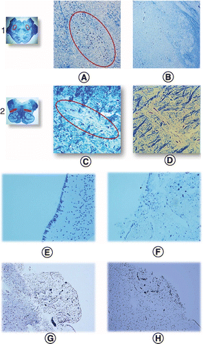
(Figure 1A–D). Interesting was the frequent observation of erosion of both the ependyma and area postrema, which are brain protective structures whose integrity can be compromised by neurotoxic substances (Figure 1E–H).

Foreign particles present in all brainstem tissue samples
With the FEGESEM, the authors identified the presence of many micro- and nano-sized foreign bodies in both the test and control groups, and their elemental chemical composition was evaluated. As carbon and oxygen elements are omnipresent in biological samples, the authors could not distinguish between elemental and oxidation state (i.e., magnesium and magnesium carbonate); nevertheless, they were focused on environmental rather than biologically present elements. Due to the higher atomic density of foreign particles, they appear brighter when compared with organic tissue.
At first, the EDS spectra of the normal brain tissue were recorded. Depending on the sampling site, there were small compositional differences, reflecting tissue specificities (rostral pons, caudal pons and medulla oblongata). After fixation, the tissues contained carbon, oxygen, sodium, phosphorus and chlorine. This spectrum (i.e., blank spectrum) was subtracted from the spectra obtained when the x-ray microprobe was focused on the foreign particles.
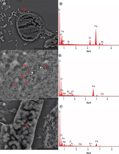
In Figure 2, in both the test and control samples, the authors identified metallic debris made primarily of iron: iron-chromium-nickel, iron-copper-silicon and iron-chromium particles. The lower part of Figure 2E is of particular interest, as it shows metallic particles inside the blood vessel, which subsequently could enter brain tissues.

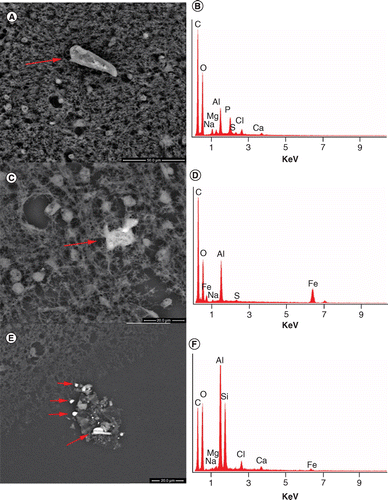
In the following figures are shown the presence of metallic particles of aluminum or aluminum-copper alloy (Figure 3) and of silver (Figure 4). Other debris of gold, titanium-silicon, nickel, zinc-copper alloy was also identified. In addition, the authors detected submicron- or nano-sized debris embedded in organic structure. These aggregates were made of biologically normal chemical constituents, only in abnormal amounts or with unusual metallic elements. The authors also noted that in the same areas where foreign bodies were identified, calcium and calcium-phosphate debris was always present. This is the ‘normal’ calcification of the brain tissue that follows inflammatory processes (as in brain metastatic calcification).


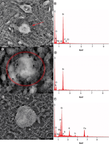
Another unexplained finding is shown in Figure 5. The pictures represent biological morphologies, but the EDS spectrum identifies a non-biological content. In fact, these structures are made of silicon. The enrichment in silicon implies a biological mechanism never described in the literature, especially not in the brain.

For a better overview, the identified foreign bodies are divided into 9 classes, according to their elemental chemical composition classification used in material science (i.e., iron-chromium and iron-chromium-nickel are two different formulations of stainless steel).
These are the classes the authors identified:
1.
Silicon [Si]
2.
Silicon-based compounds [COSiAlCaKFe]
3.
Calcium-based compounds [COCaSiAlKMgFe]
4.
Calcium compounds (as a possible result of an inflammation mechanism) [CaO, Ca–P]
5.
Al-based compound [Al, AlP, AlCu] (Figure 3)
6.
Fe compounds [Fe, FeO, FeCr, FeCrNi] (Figure 2)
7.
Silver or gold compounds [Ag, Au] (Figure 4)
8.
Titanium compounds [Ti, TiFeNiCu]
9.
Na-P-Mg compound
Actual & trending differences between SIDS/SIUDS & controls
First, despite the low number of samples, the authors were surprised to have found particles present in all the samples analyzed, including control samples (Table 3). When the data from Table 3 (number of micro- and nano-sized particles, density and age of death) were analyzed with an independent, non-parametric test, the authors confirmed that there is a trend of SIDS samples containing more microparticles than their corresponding controls (p = 0.056); SIDS controls have a generally lower number of microparticles than those found in SIUDS (cases or controls, p = 0.008); and the numbers of micro- (but not nano-) particles and their densities are statistically higher in total (case + control) SIUDS than in total SIDS (p = 0.005). In general, the authors were intrigued by the high amounts of particles found in SIUDS controls, meaning that the fetal tissue contained a higher microparticle concentration than fully grown brain tissues in already born babies.


Wow...and add the confounding variable of the rapid rise in Wireless Radiofrequency Radiation. This is also why I worry about the increase in metallic air pollution. Modern environmental illness feels quite similar to a human version of the metal spoon in the microwave. Thank you for your writing!
https://ijvtpr.com/index.php/IJVTPR/article/view/52/121