I ask myself how many people worldwide have ever thought about what could actually be in the completely useless so-called "vaccinations" and with which people are involuntarily abused as "guinea pigs" for experimental "study goals" - because various severe and also mild clinical pictures do not develop without reason - often this is accompanied by years of toxic-chemical poisoning, without the person immediately noticing/feeling anything and the organs/the organism desperately trying to detoxify on a daily basis, but it is often no longer possible - if you think about how far back in time the so-called "reference studies" of these bizarre "experiments" often go, perhaps no one should really be surprised about this anymore.....
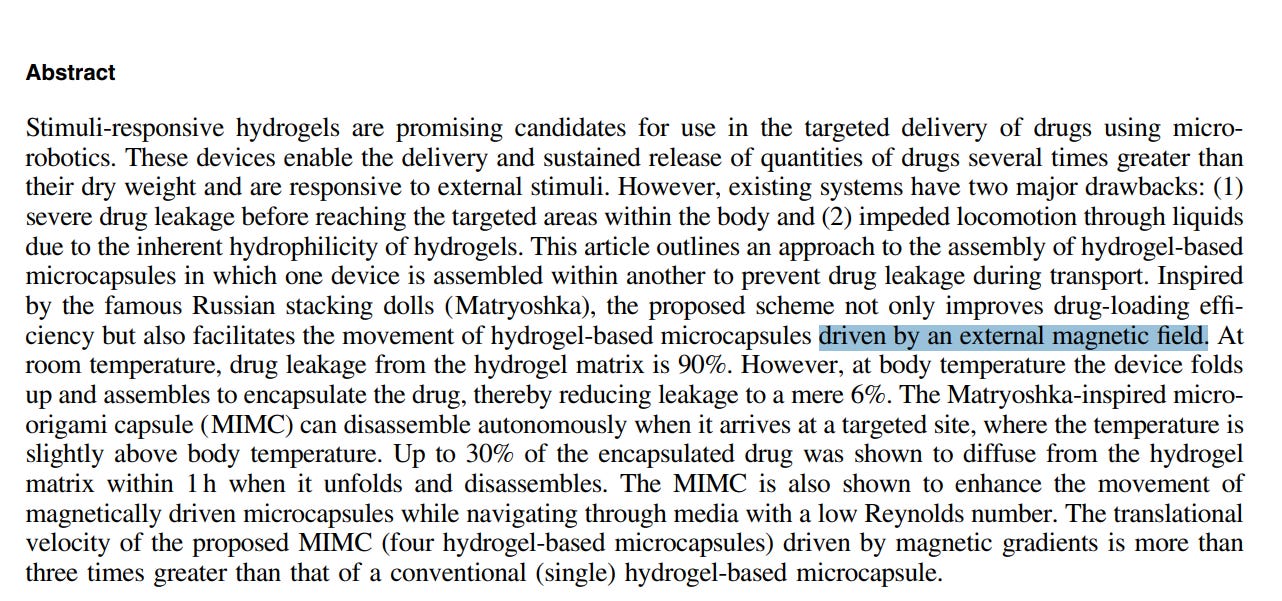
https://www.research-collection.ethz.ch/bitstream/handle/20.500.11850/306673/2/Matryoshka-InspiredMicro-OrigamiCapsulestoEnhanceLoadingEncapsulationandTransportofDrugs.pdf
https://web.archive.org/web/20240315192358/https://www.research-collection.ethz.ch/bitstream/handle/20.500.11850/306673/2/Matryoshka-InspiredMicro-OrigamiCapsulestoEnhanceLoadingEncapsulationandTransportofDrugs.pdf
Well, we know that 4 G, 5 G is part of the external magnetic field
https://ehtrust.org/scientific-research-on-5g-and-health/
and what applies to insects and animals in general is of course even more true for humans - perhaps not for everyone, but for many of them…. and we know that cells die in the process
Experiments already e.g. in 2012
https://www.ncbi.nlm.nih.gov/pmc/articles/PMC3521028/
https://www.ncbi.nlm.nih.gov/pmc/articles/PMC3521028/pdf/pone.0051085.pdf
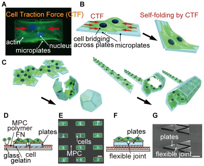
Conceptual illustration of cell origami to produce 3D cell-laden microstructures.
(A) The cells adhere and stretch across two microplates, and CTFs are generated toward the center of the cell body. Green and blue colors show actin and nucleus, respectively. (B and C) Schematic image of the cell origami: (B) the cells are cultured on micro-fabricated parylene microplates. The plates are self-folded by CTF. (C) Various 3D cell-laden microstructures can be produced by changing the geometry of the plates. (D) Schematic of the parylene microplates without a flexible joint. The cells are seeded onto the microplates coated with FN. Unwanted cells do not adhere on the glass substrate because of MPC polymer coating. (E) A fluorescent image merged with phase contrast image of NIH/3T3 cells patterned only on the microplates. The cells are bridged across the microplates. (F) Schematic of the parylene microplates with a flexible joint to achieve precise 3D configurations after folding. (G) A SEM image of the microplates with the flexible joint. Scale bars, 50 µm.
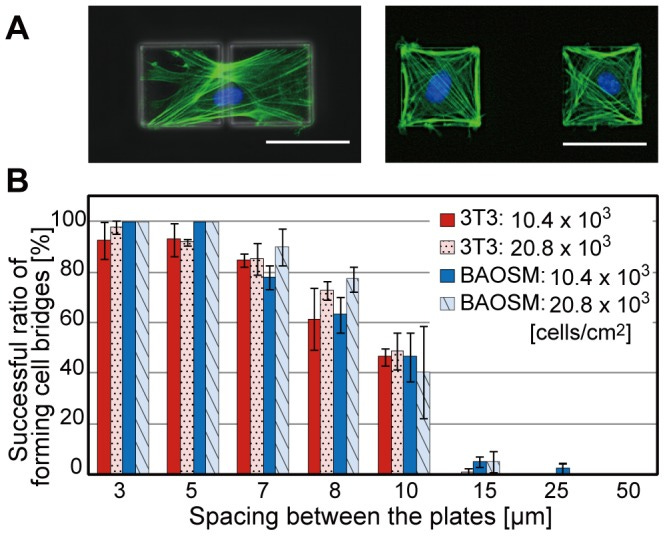
Construction of a cell bridge between a set of two microplates.
(A) Fluorescent images merged with phase contrast images of BAOSMCs on a set of two microplates with spacing of 5 µm (top image) and 50 µm (bottom image). The actin filaments and nuclei of the patterned cells on the microplates are fluorescently stained green and blue, respectively. (B) A graph of the ratio of successfully-formed cells bridge between the two microplates against various plate spacing, different concentrations of the cultured cells, and two different cell types. Results are shown as the mean ± s.d. (n = 3–10: 100 samples were observed each experiment). Scale bars, 50 µm.
Best wishes to you all from the bottom of my heart!!!♥️♥️♥️


